Advances in bioholography and digitization - Qian Li, Ph.D
2021-11-09
In the study of biomedical and life sciences, the biological cells in the glassware in the nutrient solution and under natural conditions for visual observation, expects to receive cell structure form, the dynamic characteristics, physiological parameters, the interaction between cells, cell reactions to drugs and drug transport information, such as for early diagnosis and drug design and development is of great significance.
But with the rapid development of biomedical and life sciences, these methods cannot meet the demand of dynamic quantitative observation of living cells, such as jersey Nick phase contrast microscope and differential phase contrast microscope is the basic principle of the phase information into intensity information, the transform generally is non-linear, and measurement of phase information is only qualitative. Although confocal laser microscope and optical coherence tomography can measure the three-dimensional morphology of objects, both of them require accurate scanning of samples, resulting in complex structure of imaging system and long imaging duration, which cannot achieve real-time dynamic measurement. Other improved full-field quantitative phase contrast imaging methods, such as Hilbert transform phase microscopy (HPM), Fourier transform phase microscopy (FPM) and diffraction phase microscopy (DPM), have been proposed based on the above techniques. However, some of these methods require complex mechanical motion devices such as scanning mirrors and pads, while others require wavefront sensors and multiple exposures.
Digital holography uses a planar photodetector (such as CCD, CMOS) to replace the traditional optical holographic recording medium (such as silver salt dry plate, photographic film), and stores the hologram in a digital form in a computer. Then, diffraction propagation theory is used to simulate the transmission process of light wave during holographic reproduction, and the amplitude and phase distribution of object light wave field are obtained through numerical calculation, and the whole process of hologram recording, storage and reproduction is digitized. Digital holography technology has the following advantages: 1) real-time: photoelectric image sensor can record each instantaneous state of the moving object, the reproduction process saves the tedious chemical wet processing process, improves the speed of image information processing, can be used for real-time processing occasions; 2) Rich information: a single hologram can be used to quantitatively obtain the amplitude and phase information of the reconstructed image of the recorded object, which can not only get the topography distribution of reflective objects, but also suitable for quantitative phase contrast imaging of transparent or translucent objects; 3) Simple structure: non-destructive detection of small objects can be carried out without any scanning device, and the object light wave distribution under different reproduction distances can be easily adjusted to imaging the object space layer by layer to achieve full-field observation and measurement; 4) Flexibility: modern digital image processing technology can be used to process digital holograms to reduce or eliminate the influence of aberration, noise and distortion during holograms recording and improve the quality of reconstructed images; 5) Nondestructive: digital holography does not need to be treated like fluorescence microscope to test samples for dyeing and other pretreatment. In summary, digital holographic imaging is a new real-time, non-destructive three-dimensional quantitative phase contrast imaging method with full field of view.
The lateral resolution of digital holographic microscopy (DHM) depends on the numerical aperture of the system and can be submicron, while the axial resolution can be subnanometer. And because a non-destructive and real-time digital holographic technology, particularly suitable for no stain mark quantitative phase contrast imaging of biological living cells and 3 d reconstruction, can obtain real-time cell morphology and function analysis, it will provide all kinds of quantitative parameters for biomedical research, such as the metabolism of cells, life and death, apoptosis, plasticity, signal transmission, the movement of the cell membrane and cytoplasm, Cell dynamic process and imaging of biological tissue. In recent years, it has attracted great attention from researchers at home and abroad, and has been gradually applied to the research fields of biology and medical imaging.
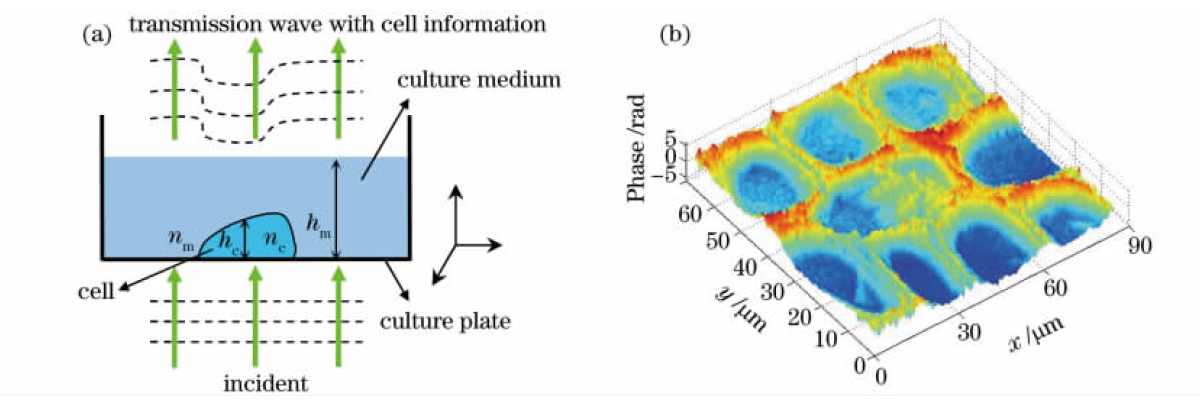
With the rapid development of digital holographic technology, digital holographic microscopic imaging has been successfully used in static cell morphology detection, biological parameters measurement and cell dynamics and other biomedical research. Based on the current research work, digital holography technology can be further combined with endoscope technology and chip technology for the diagnosis of early malignant tumors and other pathological root causes, such as viral infection, neurodegeneration and inflammation, making clinical diagnosis possible. Digital holography, combined with fluorescence microscopy and wavefront shaping technology, is expected to observe more protein expression in biological samples, realize imaging in anisotropic media and in vivo observation of cells, and then realize multifunctional microscopic system. Digital holography can also be combined with biophotonics to observe the cell function and structure at molecular level, so as to realize the detection, processing and transformation of biological system with photon and its technology. In addition, the combination of digital holography and terahertz imaging has attracted extensive attention in recent years. THZ imaging has the advantage of its opaque non-metallic and nonpolar substances (such as tumor tissues) has high penetrating power, for the visible light and near infrared light could not be achieved, and because of terahertz radiation without X ray ionization characteristics, so as not to cause harm to the material and human body, make its application in many aspects has more applications than X-ray advantage, It has a great prospect in medical examination, safety detection, environmental monitoring and space remote sensing. Its schematic diagram is shown in Figure 1.

FIG. 1 Principle of phase contrast imaging of biological samples. (a) Phase delay of incident biological cells; (b) Phase contrast of tree ring cells
Digital holography reproduction is an important part of digital holography, which is related to the success or failure of digital hologram analysis. Holograms can be reproduced by hardware using optical principles, but such a method is easy to do for a small number of holograms, but for holographic videos, it takes a lot of time to cut them and make them into holograms. Hologram reconstruction can also be realized by computer (i.e. digital reconstruction), which is more conducive to the later processing of hologram reconstruction object light wave information. Digital reproduction can be divided into two main categories: one is coaxial reproduction, the other is off-axis reproduction. The basic principle of both coaxial and off-axis reproduction is diffraction of light.
Scalar diffraction theory is mainly expressed in three formulae: angular diffraction formula, Rayleigh-Sommerfeld diffraction formula and Kirchhoff diffraction formula. These three formulas are unified in theory, but the difference is that the angular diffraction formula expresses the diffraction process of plane light wave from the perspective of frequency domain, while the latter two express the propagation process of spherical light wave from the perspective of spatial domain. The last two formulas are the general description of the expression, can not be directly used to calculate. These two formulas must be further treated if they are to have any practical value. The most common treatment is Fresnel diffraction approximation theory, which is suitable for near-field diffraction and can be implemented in two ways: Fourier transform form and convolution form. The second is fraunhoffer approximate diffraction formula, which is suitable for far-field diffraction. No matter which form of expression in scalar diffraction theory, after passing the Fresnel approximation, it can be realized by one or two forms of Fresnel approximation, which are two methods of representation -- Fresnel Fourier representation and Fresnel convolution representation. In addition, the method of using angular diffraction formula to reproduce is called angular spectral method. Whether it's angular spectrum, whether it's Fourier transform, whether it's convolution, they're all implemented by Fourier transforms, they just take different numbers of transformations.
When the digital hologram is reproduced, the reconstructed image will be clear only at its recording distance, which is also called Focus Distance (FD). When the reconstructed image deviates from this position to a certain extent (depth of field), the reconstructed image begins to blur. The larger the deviation degree is, the more blurred the reconstructed image becomes, and finally, no target can be obtained. Therefore, in order to obtain a clear reproduction image, the first step is to determine its focusing distance. The recording distance is usually not clearly known, but within a certain range when a hologram is recorded on site. If you rely on manual reproduction of digital holograms frame by frame, it will waste a lot of time.
